IN THIS POST
Skin cysts on dogs are sacs that are lined with secretory cells and which are found on the skin. Once the sacs form, there is a buildup of fluid resulting in swollen bumps on the skin. While some of the cysts will retain the fluids, others will dry out and thus the cyst will end up with firm material inside. Find out the various types of cysts on dogs including sebaceous and follicular cysts, how to drain them, as well as the procedure used to care for a burst cyst.
Cysts on Dogs
Cysts are an enclosed pocket of fluid or semi-solid material found within layers of skin tissue. The contents of the cyst may be made up of natural body fluids such as sebum (yellowish oily substance that lubricates the skin and hair of mammals), or may contain abnormal materials such as dead cells or keratin (a protein found in skin). The size can vary from 0.1 to 2 inches in diameter, but they are usually on the smaller side of that range. Cysts are usually not attached to the underlying muscle tissue and are usually painless.
Cysts can appear anywhere on the dog, but some types of cysts form in specific locations, and some dog breeds are predisposed to certain types of cysts. Below are some of the more common types of cysts that can be found on a dog’s skin.
Types of Cysts on Dogs
Cysts on dogs can be classified into different types depending on their characteristics and causes. They include:
True Cysts
These have a membrane lining the inner surface of the cyst, which produces secretions. The membrane is known as a secretory lining. True cysts mostly form as a result of blocked ducts and are common in glands. An example of a true cyst is one forming in the sweat gland, but they can also be found in other glands and elsewhere in the body besides the skin. A common location for true cysts to form is the eyelid. They cannot be prevented from occurring, but they can be removed if necessary, or the lining of the cyst can be destroyed in order to prevent recurrence.
Epidermal Inclusion Cysts, Sebaceous Cysts on Dogs
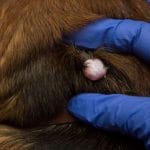
Epidermal inclusion cysts, also known as epidermoid cysts, follicular cysts, or sebaceous cysts, are the most common type of cysts in dogs. These occur when the top of the hair follicle becomes covered with epithelial (skin) cells and keratin, a skin protein, is secreted into the newly formed cyst. The material within the cyst is either fluid or a dark-colored cheesy substance; the cyst itself is usually firm and round in shape. Comedones, or blackheads, are a similar type of cyst, but they have a wide opening at the surface of the skin. Sebaceous cysts are formed by the blockage of the sebaceous glands, which are located around hair follicles, and contain sebum, which is a waxy substance that coats the skin and hair.
Epidermal inclusion cysts can be caused by injury to the skin, blockage of the pore or hair follicle, sun damage, pressure point damage, or lack of activity in the hair follicles such as with Mexican hairless or Chinese crested dogs. Boxers have a predisposition for getting multiple and recurrent epidermal inclusion cysts on their heads. Schnauzers, shih tzus, basset hounds, and Yorkshire terriers also have an inherited predilection for epidermal inclusion cysts. Comedones can form on the sternum and other pressure points in breeds with a thin haircoat and little body fat, such as the greyhound. Sebaceous cysts tend to form on the head, neck, torso, or upper legs.
These types of cysts are prone to secondary infection, and as tempting as it might be, they should not be squeezed or popped. The keratin contained within the cyst could be pushed deeper into the surrounding tissue, causing a severe localized immune response. The implosion from a squeezed cyst could also lead to bacterial cellulitis, an infection of the tissues underneath the skin, which would need to be treated with antibiotics.
Interdigital Follicular Cyst on Dogs
Interdigital follicular cysts in dogs are quite common. They usually appear as blood- or pus-filled red nodules between the toes, usually on the front feet, and form as a result of excessive friction or trauma to the webbing between the toes. There are many factors that may be involved in the formation of these cysts such as a short and coarse hair coat, wide paws, obesity, difficulty walking such as due to arthritis, and excessive licking and chewing of the feet. Breeds that are especially susceptible to interdigital cysts include boxers, pit bulls, and Doberman pinschers.
Interdigital cysts on dogs can be treated with surgery, CO2 laser, or medical therapy. Surgery tends to be the least recommended option because the affected webbing is removed and the toes are fused together; this can lead to orthopedic issues in the future because the structure of the paw is changed. CO2 laser removes the cysts from the webbing by vaporization, which allows the webbing to heal with normal skin tissue, but multiple treatments may be needed for severe cases. With medical therapy, either oral or topical steroids are used to reduce inflammation, and antibiotics are used to clear up the infections that commonly occur with interdigital cysts.
Dermoid
Dermoid cysts are congenital (present from birth) cysts that form along the top midline of the head or along the spinal column. These malformations differ from other cysts in that they contain fully formed hair shafts due to the infolding of the skin during fetal development. While dermoid cysts are rare, they are most commonly seen in boxers, Rhodesian ridgebacks, and Kerry blue terriers.
False Cysts
These are fluid-filled sacs but which do not have the secretory lining found in true cysts. False cysts develop in response to trauma or hemorrhage that leads to tissue death and are filled with fluid. The fluid comes from the liquification of the dead tissues. They are fairly common in dogs, especially when the injury occurs along the flanks (area along the side between the ribs and the hips). Rarely, these false cysts will form in reaction to an injection.
Diagnosis of Cysts on dogs
While cysts are generally benign (not harmful), it can sometimes be difficult to distinguish a benign cyst from a malignant skin tumor, so consultation with your veterinarian is recommended, particularly if you notice any of the signs listed below:
- Rapid growth
- Discharge or oozing blood
- Changes in shape
- Color changes
- Textural changes such as from soft to hard
- Any other change in appearance
Usually the first step in diagnosis is for your veterinarian to perform a fine needle aspirate (FNA) of the lump. Cells are removed from the lump using a needle attached to a syringe. The cells are then placed on a microscope slide, stained, and then examined under a microscope. Your veterinarian will usually be able to give you a diagnosis within a few minutes, but it may be necessary to send the sample out to a laboratory for a veterinary pathologist to review the slide and provide a definitive diagnosis. One downfall of the fine needle aspirate is that the veterinarian might not be able to collect enough cells. Another potential problem is that since the lump may have several different types of cells present, not all of the different types of cells may be collected, leading to potentially missing the cell type that would provide the definitive and correct diagnosis.
If there is any uncertainty with the fine needle aspirate, your veterinarian may recommend a tissue biopsy or perhaps even an excisional biopsy. With a tissue biopsy, your dog is administered a local anesthetic, and a small piece of the lump is removed and sent to the laboratory for analysis. While a definitive diagnosis is very likely with this approach, a drawback is that if the pathology results indicate a malignant growth, another procedure will have to be performed to remove the growth.
An excisional biopsy is performed while your dog is under general anesthesia and involves the removal of the entire lump and some of the surrounding tissue to ensure that all of the lump has been removed. This approach will yield a definitive diagnosis, and if it turns out to be a malignant growth, the pathologist will also comment on whether they believe all affected cells have been removed from the surgical site. However, it is the most invasive and expensive approach, and the growth may turn out to be benign, meaning the procedure may not have been necessary.
Each diagnostic method has its pros and cons. Your veterinarian will make a recommendation based on the appearance, location, and size of the growth, and together you can decide what makes the best sense for your dog.
Treatment
Uncomplicated cysts are harmless and usually only need treatment when they are too unsightly or to the point of inhibiting your dog’s movement or causing discomfort. In cases where treatment is not necessary and they are left to be, one of three likelihoods will occur:
-
They will disappear with time
-
They will burst and drain off the contents before healing
-
They will remain as lumps beneath the skin without growing or disappearing.
If treatment is necessary, it is usually accomplished by surgical removal of the cyst. This procedure is performed under general anesthesia. The surrounding area will be clipped and scrubbed, the cyst is surgically removed, and sutures are placed in the skin. Antibiotics and pain medications will be sent home for use after the surgery. Your dog may also be sent home with an E-collar to prevent him from disturbing the sutures.
Right after a dog’s cyst removal, the results can be seen immediately. The symptoms associated with it such as swelling should start fading away.
How to Treat a Ruptured Cyst
Sometimes the cysts will rupture on their own. When this happens, it is important to keep the area clean to prevent secondary infections as well as promote fast healing. Here is how to clean a ruptured cyst on a dog:
-
If necessary, trim the area around the cyst to gain access and allow for direct contact with the cyst. Trimming will also prevent the discharge from crusting on the fur.
-
Using a damp but warm washcloth, clean up the area. Start by applying it as a warm compress until the wash cloth loses heat. This will help to drain the cyst further as well as soften any crusting that may have formed from the contents of the cyst for ease of removal.
-
Follow this up by disinfecting the area. You can use a product like Vetericyn Plus, or hydrogen peroxide.
Repeat this procedure until the area heals completely. Keep monitoring the skin around the ruptured cyst since ruptured or burst cysts are vulnerable to infection. Inflammation and itchiness are also quite likely to occur. Your dog should be prevented from licking at or scratching at the ruptured cyst; it may be necessary to get an E-collar to help keep them away from the wound. If you notice any swelling, redness, discharge, warmth to the touch, or odor, take your dog to your veterinarian for examination and treatment.
Related posts:
 Sebaceous Cyst on Dog: Removal and Care When Ruptured, Bleeding
 Bumps on Dogs Skin, Back, Head, Chin, Face and Nose – Treatment Options
 Bald Spots on Dog (Tail, Leg, Back): Causes & Treatment
 Dog Pimples – Causes, Treatment, and Pictures
 Dog Rash on Belly: Causes and Treatment
 Pale Gums in Dogs: Causes and Treatment