A bump on your dog’s eyelid can be worrisome and scary-looking, especially if it seems to be bothering your dog. However, most bumps and inflammation are easily fixed with the proper diagnosis and treatment. Below we’ll cover the various eyelid bumps and swellings that can be seen in dogs, their diagnosis, and their treatment.
Bump on Dog Eyelid
A bump on dog eyelid may appear as a lump, growth or swelling on the eye lid surface or along it’s edges. Causes may include bacterial, viral or fungal infections in which case the bumps vanish once the infection is addressed. In some cases, bumps may be due to tumor formation and treatment can be far more involved.
Chalazion
A chalazion is a firm, non-cancerous and non-painful swelling of a meibomian gland caused by the accumulation of its secretions due to blockage of the duct. Meibomian glands are found along the edges of both eyelids; these are specialized sebaceous glands that secrete an oily substance that helps keep the tear film in place over the eye itself. Chalazia are often referred to as cysts, and are commonly seen in older dogs. Diagnosis is fairly straight-forward and based on the appearance and location of the lump. Treatment is also fairly straight-forward. With well-behaved patients, the chalazion can be drained by making an incision in the chalazion while the patient is under light sedation with local anesthesia; cryosurgery (the use of intense cold to destroy unwanted tissue) may also be used to prevent recurrence. A topical antibiotic with steroid may also be used to help reduce inflammation and prevent infection while the incision heals.
Hordeolum (stye)
A hordeolum, or stye, is an infection at the root of the eyelash. While a hordeolum is a firm and non-cancerous nodule like a chalazion, it is quite painful. Styes tend to be smaller than chalazia, and look similar to a pimple. The diagnostic hallmark of a stye is pain upon manipulation. Treatment includes drainage of the stye, topical antibiotic ointments, and warm compresses of the eyelid several times a day.
Infectious (bacterial) blepharitis
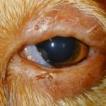
Bacterial blepharitis (inflammation of the eyelid) is usually caused by Staphylococcus or Streptococcus species. The eyelids are diffusely inflamed, red, and irritated, with discharge and crusting around the eyelid. If the inflammation is chronic, the eyelid margins may be ulcerated and exhibit hair loss. The meibomian glands may also become impacted and abscessed. Both eyes are usually involved but not always.
One method of diagnosing bacterial blepharitis is via an impression smear – this is accomplished by pressing a microscope slide against the eyelids and examining the slide under a microscope. If it’s a bacterial blepharitis, the slide will contain sphere-shaped bacteria (cocci) and neutrophils (a type of white blood cell important in fighting infections). Another diagnostic test is to grow a culture of any material expressed from an ulcer or abscess followed by antibiotic susceptibility testing to determine the best antibiotic to use to treat the blepharitis. Treatment is with oral antibiotics, usually for at least three weeks. If the inflammation is severe, oral or topical steroids may also be used. Improvement is usually seen within 7-10 days of starting treatment.
Parasitic blepharitis
Inflammation of the eyelids can also be caused by external parasites, such as those mites that cause demodectic mange and sarcoptic mange.
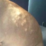
Demodectic mange is caused by Demodex canis, a cigar-shaped mite that lives in the hair follicles. Some of those mites can always be found on your dog, but as long as they have a mature and healthy immune system, they do not become infected. In puppies younger than about 10 months of age, demodectic mange can appear in a localized area around the eye or eyes. The lesions caused by mange include hair loss around the eye, mild redness and irritation, and scaling of the skin. There is often a secondary bacterial infection associated with the lesions, and this can cause swelling around the eye and a moist discharge. Older dogs with demodectic mange tend to have patches of hair loss over the entirety of their body.
Diagnosis of demodectic mange is performed by plucking some hairs from alongside the lesions on the dog’s body and then examining the hair roots under a microscope. Since the mites live in the hair follicles, they should be attached to the hair roots when the hairs are plucked. Skin scraping is also a possibility, but since the mites are in the hair roots, the scrape must be quite deep and therefore will be quite painful and take time to heal. As far as treatment goes, most localized demodectic mange will resolve on its own as the young dog’s immune system matures, but amitraz (Mitaban), ivermectin, and moxidectin can all be used for treatment of systemic disease. Amitraz is applied as a bath or dip and is usually performed by a veterinary professional. Ivermectin or moxidectin can be found in most monthly parasite preventatives. Oral antibiotics will also be prescribed if a secondary bacterial infection is present.
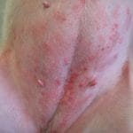
Sarcoptic mange (also known as scabies) is caused by Sarcoptes scabiei, an oval-shaped mite that burrows just beneath the top layer of skin. The burrowing and tunneling as the mites move under the skin causes intense itching, which leads the dog to scratch, bite, and chew at himself, causing hair loss and irritation to the skin. Around the eyes, the eyelids will be crusty, be thickened, and exhibit some hair loss, but with sarcoptic mange, it is unlikely for only the eyelids to be affected. Typically, the ear margins, abdomen, and elbows are affected. Sarcoptes is highly contagious and easily transmitted from dog to dog or even dog to human.
Diagnosis of sarcoptic mange is based on clinical signs, skin scraping, or response to therapy. With skin scraping, the skin is scraped with a dulled scalpel blade coated in mineral oil; the oil is then placed on a slide and examined under a microscope. Unfortunately, mites are not always found with this method, and veterinarians will sometimes treat for scabies and look for a response. Treatment is with topical selamectin (Selarid, Revolution) or moxidectin (Advantage Multi). Any bedding your dog uses should either be discarded or washed frequently with diluted bleach. Routine use of flea and tick preventatives will usually prevent sarcoptic mange infestation.
One case of Cuterebra infestation of the conjunctiva (the lining of the inside of the eyelids) has been reported in a puppy. Members of the Cuterebra genus are known as bot flies or warble flies, and generally infest rodents, but can also sometimes be found in dogs and cats. The flies are harmless, but they lay their eggs near the entrances of rodent burrows, where they are usually picked up by the rodents as they pass through. Dogs that sniff at burrows can also pick up the eggs in their fur. Once the eggs are picked up by the dog, they burrow into the skin and mature into larvae over the course of several weeks before they wiggle out of the skin and become adult flies.
Diagnosis of a Cuterebra infestation is fairly easy as there will be a lump under the skin (or in the eyelid in this case) with an opening to the outside; if you watch the opening for a minute or two, you will see the end of the larvae come to the opening in order to breathe. Removal of the larvae must be done by a veterinarian; if not done properly, the larvae can burst and cause an allergic reaction, up to and including anaphylactic shock. Your veterinarian will apply a local anesthetic, enlarge the opening slightly, use forceps to carefully remove the larvae, and then flush out the wound. Your dog will go home with oral antibiotics and pain medications, and a full recovery would be expected. There is no real prevention for Cuterebra infestation, other than preventing your dog from sniffing at rodent and rabbit burrows.
Immune-mediated blepharitis
While generally rare, several immune-mediated conditions can cause inflammation, ulceration, crusting, or scaling of the eyelids, such as pemphigus, discoid lupus erythematosus (DLE), uveodermatologic syndrome. Often with these conditions, other skin around the face is also affected, and is especially obvious around the nose. For diagnosis of these conditions, a skin biopsy is performed and sent to an outside laboratory for examination by a veterinary pathologist. Treatment is generally started with either oral or topical steroids for immediate relief, and continued with immunosuppressive drugs such as azathioprine or cyclosporine.
Viral papilloma (warts)
Warts are benign tumors of the skin caused by the papilloma virus; veterinarians refer to dog warts by the more formal terms “papilloma” or “viral papilloma”. These tumors have a classic fimbriated appearance, which means they are round, but have a rough surface similar to a sea anemone or a cauliflower. They typically occur on the lips and around the muzzle of a young dog (usually less than two years of age). Rarely, these papillomas can be found on the eyelids or between the toes. Diagnosis of papillomas on the face is usually fairly straightforward since they have a rather unique and characteristic appearance. Most papillomas in younger dogs will spontaneously disappear within 1-2 months as the developing immune system learns how to deal with the virus. Prevention of viral papillomas consists of not allowing your dog to play with dogs that have visible papillomas. You should also prevent your dog from playing with the infected dog’s toys, or using his bowls or bedding. If your dog has wounds or rashes that compromise the integrity of his skin, it’s best to keep him away from places where other dogs congregate, such as dog parks or doggy day care. And if your dog does develop papillomas, keep him away from other dogs until the tumors regress.
Tumors
There are many different types of tumors that can affect the eyelid and cause either localized or diffuse irritation of the eyelids. Most tumors affecting the eyelid are benign, affect the upper eyelid, and occur in dogs older than 10 years of age. The following list includes the more common eyelid tumors found in dogs:
-
Meibomian gland adenoma: This slow-growing benign tumor is the most common of all eyelid tumors. They arise from the conjunctiva of the eyelid and have a lobed appearance.
-
Meibomian gland adenocarcinoma: Malignant tumor that is indistinguishable from an adenoma by sight. While technically malignant, it behaves similarly to the benign adenoma.
-
Melanoma: There are two types of melanoma – the first arises from the eyelid skin and produces a single, smooth, protruding mass while the second arises from the pigmented eyelid margin, is flat and broad, and tends to expand in all directions. Both types are usually benign.
-
Histiocytoma: Benign tumor of young dogs that has a characteristic appearance of being raised, less than ⅜ inches in diameter, pink in color, hairless, and has a rapid growth pattern. These tumors generally disappear on their own 3-5 weeks after making an appearance.
Diagnosis of the specific type of tumor can sometimes be done with a fine needle aspirate (FNA) wherein a small amount of cells from the tumor are suctioned out using a needle and syringe and then examined under a microscope. However, if surgery is being planned to remove the mass regardless of the exact diagnosis, then waiting until the day of surgery and sending the entire mass out for a pathologist report could be the more reasonable course of action.
If the tumor is not causing any eye irritation or otherwise interfering with the dog’s sight, then it may be reasonable to take a wait-and-see approach. However, if that is not true, or if the mass has the potential to be more difficult to remove as it grows larger, then surgical removal of the mass is the prescribed treatment. Depending on the size and exact location of the mass, your veterinarian may refer your dog to a veterinary ophthalmologist due to the delicate nature of some surgeries and the potential need to reconstruct a functioning eyelid. Cryosurgery (using extreme cold to destroy unwanted tissue) may also be used to hopefully prevent recurrence of the tumor.
Take away message for dog eyelid bumps
There are many causes of either bumps or inflammation of your dog’s eyelids. Many are easily diagnosed and treatable by your regular veterinarian, but some conditions require referral to a veterinary ophthalmologist. Since your dog’s eyesight is so important to protect, be sure to have your dog examined by your veterinarian when you see anything around your dog’s eyes that is cause for concern.
- https://groommypets.com/bump-on-dog-eyelid/