IN THIS POST
Dizziness in dogs is likely due to a malfunction of their vestibular system. The vestibular system is what tells your dog where his head and body are in space, and which direction his body is turning. There are two components to the vestibular system: the peripheral system which is located in the inner ear, and the central system which is located in the brain. Conditions affecting the peripheral vestibular system are generally benign or easily treated, while conditions affecting the central vestibular system are more serious. Your veterinarian may refer your dog to a veterinary neurologist for workup and possibly an MRI to determine the cause of the vestibular symptoms. Since an MRI can be or will be recommended for all of the causes of vestibular dysfunction, we’ll discuss this important diagnostic tool in detail before discussing the potential causes.
Dizziness in dogs, Veterinary neurologists and the MRI
A veterinary neurologist is a veterinarian who completed a year or two of internship in internal medicine or neurology and then a three year residency program in neurology after completing their four year veterinary degree. They also need to publish at least one original research study and pass two intense days of testing before becoming a boarded neurologist, also known as a Diplomate of the American College of Veterinary Internal Medicine (DACVIM (Neurology)).
One of the most common diagnostics offered at a neurologist is an MRI (magnetic resonance imaging) scan, though a CT scan (computerized tomography; a 3D X-ray) may also be able to provide the images required for a diagnosis, depending on the suspected cause. A CT scan, since it uses X-ray radiation to produce the images, is excellent for visualizing bone. However, an MRI is superior to a CT in providing images of soft tissue, such as the brain and spinal cord, thus making it the advanced imaging choice of neurologists.
Instead of X-ray radiation, an MRI uses magnets to align the water molecules of the body in one direction and then sends a pulse of radiofrequency (RF) energy to knock certain water molecules out of alignment. The water molecules return to alignment with the magnet after the RF pulse ends, and that movement gives off energy that is detected by a receiver (“coil”) placed around the patient. That energy signal is then processed by a computer to produce a detailed image of the part of the body being scanned. Different types of images can be produced by varying the sequence and timing of the RF pulses, and the different types of tissue (cerebrospinal fluid, white and gray matter of the brain, fat, infection/inflammation, etc.) will show up as either light or dark on the MRI image depending on the RF pulses used to produce the image. The neurologist knows what a normal scan looks like and will be able to pick out any changes from normal, even changes as small as 1-2 millimeters in size.
The MRI machine used is the same as used in human medicine. However, in order for usable images to be collected, the patient must lie absolutely still for the entire time of the scan. Scans of the head take about 45 minutes to complete, and it is very noisy in the MRI suite, so dogs are placed under general anesthesia for the scan, both for keeping them still and also preventing them from being frightened and stressed by the noise. Dogs undergoing an MRI scan will have earplugs placed in their ear canals to prevent damage to their hearing. Usually two people perform the scan under the guidance of the neurologist – one to program and run the MRI scan and the other to monitor your dog’s vitals while under anesthesia. Results of the scan are read in real time by the neurologist, but for unusual scans, another neurologist or radiologist may be consulted via telemedicine. Depending on the severity of the condition found on the scan, you may be called while your dog is still in the scanner, but most often, you will meet the neurologist and review the results of the scan when you pick up your dog later in the day after he has recovered from anesthesia.
Now that we know more about veterinary neurologists and the MRI scan, let’s take a look at the various conditions that can cause vestibular dysfunction and how neurologists and advanced imaging can help with the diagnosis of the underlying cause of vestibular dysfunction.
Peripheral vestibular system, Dizziness in dogs

The peripheral vestibular system consists of the semicircular canals of the inner ear and the vestibulocochlear nerve that runs through the inner ear and transmits information to the vestibular system located in the brainstem and cerebellum of the brain. Symptoms of peripheral vestibular problems include head tilt, loss of balance, leaning, falling, or rolling to the side, vomiting/nausea, facial paralysis, and a twitching of the eyes called nystagmus. Conditions that cause peripheral vestibular dysfunction are generally benign and have a good prognosis for recovery with appropriate treatment. However, it is likely that dogs affected with peripheral vestibular syndrome will have at least some degree of a head tilt the rest of their lives; this does not affect their quality of life.
Idiopathic vestibular disease (“old dog” vestibular disease)
While many causes of vestibular disease are understood and able to be characterized, sometimes vestibular symptoms are idiopathic, which means they appear spontaneously without any apparent cause. Idiopathic vestibular disease in dogs appears to be related to age (the average age of onset is about 12 years old) thus it is usually referred to as “old dog vestibular disease”. Idiopathic vestibular disease is the most common type of vestibular disease, making up 68% of cases reported in a recent study.
Symptoms appear suddenly and may worsen over 12-24 hours, but your dog will start to recover on his own within 24 hours of symptom onset. If he continues to worsen after 24 hours, then it is unlikely to be idiopathic vestibular disease, and the cause should be investigated. Diagnostics include an examination, blood work, and advanced imaging (CT or MRI).
Unfortunately, since there is no known cause for old dog vestibular disease, there are no medications or treatments that can be given to address the cause and speed recovery, nor is there anything to be done to prevent its occurrence.
Anti-nausea medications such as Cerenia (prescription only) or meclizine (available over the counter as Bonine or non-drowsy Dramamine) can be given to alleviate any nausea, vomiting, or motion sickness your dog may be experiencing. Symptoms will gradually resolve over the course of one to two weeks. Recurrences of idiopathic vestibular episodes are uncommon but not impossible.
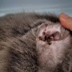
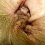
Otitis interna
An infection of the inner ear is another common cause of peripheral vestibular disease, accounting for about 26% of all cases, particularly in younger dogs. The head tilt and loss of balance will be towards the side with the infected ear, unless both ears are infected. In that case, the dog will usually swing his head back and forth from left to right and may lean or fall to either side.
Diagnostics include an examination of the ear canal, and in particular, an attempt to see the eardrum and determine if that has been ruptured, allowing bacteria to make its way into the inner ear. Other diagnostics can include advanced imaging (CT scan or MRI) and bacterial cultures of mucus present in the ear as a result of the infection. The imaging allows the neurologist to see if the infection has crossed over into the brain (making it a more serious situation), and if it has, a spinal tap may be recommended. Both the imaging and spinal tap are done under general anesthesia, with the spinal tap immediately following the scan. The spinal tap is used to collect a sample of cerebrospinal fluid, the fluid that surrounds the brain and spinal cord. The fluid is then analyzed to determine if an infection or inflammation has indeed spread to the spinal fluid and is causing a meningitis. The bacterial culture of the material in the middle/inner ear is sent out to a veterinary laboratory, where they will attempt to grow the bacteria, and if successful, test that bacteria against various antibiotics to determine which antibiotic would work best against the bacteria that is present in the inner ear.
Treatment for otitis interna is a course of oral antibiotics for a minimum of six weeks. The inner ear is buried deep within the bones of the skull, making it difficult for antibiotics to easily reach the infection. Depending on the severity of the dizziness and consequent nausea, a brief hospital stay with IV fluids may be recommended in order to maintain hydration until the nausea subsides. Anti-nausea medications may also be prescribed. If the symptoms do not improve within three weeks, or if they get worse, then an ear canal flushing procedure called a myringotomy may be recommended; a veterinary dermatologist is the best person to perform the procedure. The vestibular signs should resolve as the infection clears, but any head tilt that was present may never completely resolve.
While it’s not completely possible to prevent inner ear infections, chronic or severe outer ear infections predispose your dog to an inner ear infection, so careful monitoring for and prompt treatment of ear infections (otitis externa) can help reduce the chances of an inner ear infection occurring.
Hypothyroidism
Hypothyroidism, or low thyroid hormone, can cause vestibular symptoms through mechanisms that are poorly understood, and account for about 4% of cases. Affected dogs may have other signs of hypothyroidism such as weight gain, lethargy, hair loss, slow heart rate, and weakness.
Diagnosis of hypothyroidism is achieved by a blood test. Usually, measuring the level of T4 (thyroxine, a thyroid hormone), is sufficient for a diagnosis of hypothyroidism, but some dogs, and sighthounds in particular, normally have low thyroid hormone levels but are not considered hypothyroid. Therefore, it may be best to have a full thyroid blood panel (T4, fT4, and cTSH at a minimum) performed in order to diagnose hypothyroidism, particularly in sighthounds such as greyhounds, borzoi, Afghan hounds, deerhounds, and whippets.
If your dog is diagnosed with hypothyroidism, he should be supplemented with thyroid hormone, which is in the form of a small tablet given by mouth every 12 hours. Vestibular signs are usually reversible once thyroid supplementation has begun, but it may take up to four weeks for the signs to completely resolve.
Neoplasia
While there are some tumors that affect the inner ear, these are fairly rare, and account for less than 1% of all peripheral vestibular dysfunction cases. The most common tumors that can invade the middle and inner ear are ceruminous gland adenocarcinomas and squamous cell carcinomas.
Recommended diagnostics include advanced imaging as well as a biopsy of the tumor if it is readily accessible from the outer ear. Depending on the type of tumor, surgical debulking (removing as much of the tumor as possible), radiation therapy, chemotherapy, or palliative care may be recommended. Prognosis is generally guarded to poor.
Congenital peripheral vestibular disease
Some breeds have a predisposition to vestibular dysfunction caused by degeneration of the inner ear structures or a malformation of those structures. Symptoms usually appear around 3-4 weeks of age (when the puppies first begin to really walk and move around). Many puppies learn to compensate by 2-4 months of age, but some will continue to be affected to some degree the rest of their lives. Most affected puppies will also be deaf by 3 weeks of age due to the degeneration of the inner ear.
Diagnosis is made by physical examination, age, breed, and hearing evaluation. The most commonly affected breeds include doberman pinschers, German shepherds, English cocker spaniels, and beagles. An evaluation of the ability to hear is often difficult to perform due to the puppy’s compensation for the hearing loss so a special hearing test called a BAER (brainstem auditory evoked response) test may be recommended. This test involves measuring the brain’s electrical response to sound, similar to how an EKG measures the electrical activity of the heart. A BAER test is performed under the supervision of a veterinary neurologist.
There is no treatment for congenital peripheral disease. Affected puppies should have their hearing tested and if found to be deaf, training the dog to respond to visual commands should be started immediately. The affected dog, his parents, and his siblings should not be allowed to breed in order to prevent the genetic mutation from continuing to spread in the population.
Dizziness in dogs, Central vestibular system
The vestibulocochlear nerve that runs through the inner ear sends information about head and body position to the brainstem and cerebellum. The portions of the vestibular system located in the brainstem and cerebellum comprise the central vestibular system. Symptoms of central vestibular dysfunction are very similar to peripheral vestibular dysfunction: head tilt, loss of balance, rolling or falling to one side, and/or nystagmus (rhythmic twitching movement of the eyeballs).
However, dogs with central vestibular dysfunction usually also have other neurologic deficits. For instance, they usually do not know where their paws are in space. This is called a postural deficit and is tested by flipping your dog’s paws over so they are standing on the tops of their paws. Dogs that are normal or have only peripheral vestibular dysfunction will immediately flip their paws back over to the correct position, whereas dogs whose central vestibular system is affected will not flip their paws back over the vast majority of the time. Patients with central vestibular dysfunction may also have a change in mentation, meaning that they are dull or in a stupor. Veterinarians will also assess function of the nerves affecting the head (cranial nerves); dogs with central vestibular signs commonly have reduced facial sensation, reduced gag reflex, and/or reduced tongue movement. Since the brain is involved, the underlying cause of the vestibular signs is generally more serious and the overall prognosis more grim compared to peripheral vestibular causes.
Neoplasia
Tumors located in the cerebellum or brainstem can cause vestibular signs. It is also possible that a tumor located in the forebrain can grow large enough to place pressure on the cerebellum and/or brainstem and cause vestibular signs, but this is far more common in cats versus dogs. The most common tumors that arise in the cerebellum or brainstem include gliomas, meningiomas, and choroid plexus tumors.
An MRI is used for diagnosis of the tumor and also to determine its exact location in the brain. Surgical excision of the tumor may be possible if it is accessible. Otherwise, radiation therapy, chemotherapy, or palliative control of brain swelling using steroids are the recommended treatments (depending on the tumor type). Various clinical trials are usually also available; ask your veterinary neurologist if one is available near you.
With the exception of meningiomas, there aren’t any large-scale studies in the veterinary literature regarding average survival time, but the prognosis is considered to be poor. With regards to meningiomas, average survival times range from 7 months for surgery or radiation therapy alone to 2 years with surgery followed by radiation therapy.
Encephalitis/meningoencephalitis
Encephalitis is an inflammation of the brain whereas meningoencephalitis is inflammation of the brain and the meninges, which consist of three layers of membranes that surround and protect the brain and spinal cord. There are a number of possible causes for inflammation of the brain and/or meninges; these range from immune-mediated to infectious causes due to bacteria, fungi, protozoa, or fungi. The most common form of immune-mediated encephalitis is granulomatous meningoencephalomyelitis (GME), which is most often reported in young adult toy and small breeds, though it can occur in any breed at any age. The most common form of infectious encephalitis is caused by the canine distemper virus; affected dogs are usually less than one year of age, though dogs that are affected with central vestibular signs are usually at least a year old.
Encephalitis can affect multiple areas of the brain (otherwise known as multifocal or disseminated disease), so in addition to central vestibular signs, seizures, behavior changes, circling, pacing, and acute blindness may also be seen. Diagnostic tests include an MRI and spinal tap. If an infectious cause is suspected, then additional testing to determine the infectious organism can be performed on blood and also the cerebrospinal fluid (CSF) collected during the spinal tap.
Treatment of immune-mediated encephalitis (GME) is directed at dampening the immune system. This is achieved by administration of a chemotherapy agent called cytarabine (Cytosar), which is usually given as an intravenous constant rate infusion (CRI) over 24-40 hours while your dog is hospitalized. Intravenous drugs such as mannitol or hypertonic saline are given to reduce swelling of the brain, and steroids are started, both to help reduce inflammation and also to suppress the immune system. When your dog goes home, he will usually be prescribed a steroid and an oral immunosuppressant drug called cyclosporine in order to prevent relapse. He will likely be on at least one of those drugs for the remainder of his life.
Prognosis for recovery from GME is guarded, with approximately one third of dogs dying or being euthanized due to poor quality of life within the first week after diagnosis. Of those that do survive the first week, average survival time for GME has been reported as less than 18 months, but this is based on older research studies that are skewed due to several factors. More recent anecdotal evidence suggests that survival time is usually well over 2 years after diagnosis, assuming the patient survives the initial illness.
Treatment of encephalitis due to infectious causes will depend on the infecting organism. Antibiotics are used for bacterial infections while antifungals like fluconazole are used for fungal infections. No medications are available to treat viral infections such as canine distemper; instead supportive care is provided while the dog’s immune system fights the disease. Prognosis for infectious encephalitis is generally guarded to poor, though the prognosis for bacterial or protozoal causes may be better if the disease is diagnosed early and treated aggressively.
Infarction
Until recently, infarcts, or strokes, were believed to be uncommon in veterinary medicine. But with greater availability of MRI in veterinary neurology, it has become apparent that strokes are more common than originally thought. Strokes that cause central vestibular signs generally occur in the cerebellum versus the brainstem, and tend to occur in small breed dogs and greyhounds. There are several underlying causes of infarction; these include high blood pressure, hyperadrenocorticism (Cushing’s disease), chronic kidney disease, protein losing nephropathy, hypothyroidism, cardiac disease, or coagulopathies (von Willebrand, hemophilia).
Diagnosis requires an MRI; the infarction becomes visible on MRI 12-24 hours after the onset of symptoms. If a stroke is suspected based on MR imaging, tests to determine the underlying cause of the stroke are recommended. These tests may include serial blood pressure monitoring, thyroid level, coagulation profile, echocardiogram, and/or urinalysis with urine protein:creatinine (UPC) ratio. A basic blood work panel should have been performed before anesthesia for the MRI; this panel would help screen for kidney disease and Cushing’s disease as possible causes.
Treatment for an infarction is generally supportive and focuses on making sure that the brain is maintaining an adequate blood supply. Hospitalization may be needed for the first day or two in order to monitor your dog and provide IV fluid support in order to maintain appropriate blood pressure and thereby appropriate blood supply to the brain. If an underlying cause can be identified, then treatment of that underlying condition should help prevent future recurrences.
The prognosis is variable, and depends on the degree of infarction and the severity of any concurrent illnesses. Many dogs will make a partial to full recovery with enough time and supportive care. While it’s not possible to entirely prevent strokes from occurring, the risk can be reduced by working with your veterinarian to monitor your dog’s blood work and health at least once a year, and ideally twice yearly, as your dog becomes a senior.
Metronidazole toxicity
Metronidazole (brand name Flagyl) is an antibacterial and antiprotozoal drug used in treating some GI infections, and may also be used to help treat inflammatory bowel disease. Metronidazole is generally very safe at appropriate dosages, but can be toxic at very high doses given over a short period of time, or at moderately high doses given chronically. Central vestibular signs of being off-balance, nystagmus, and postural deficits are present in metronidazole toxicity along with nausea and vomiting.
Diagnosis is fairly straightforward – if your dog is exhibiting these symptoms while taking metronidazole, particularly at higher doses or long-term, the drug is likely the cause. Stopping the drug will result in resolution of the symptoms, but it may take a week or two for the signs to disappear. However, diazepam (tranquilizer, controlled drug) has been shown to reduce the time to resolution of symptoms from 14 days to as little as 3 days, so depending on the severity of symptoms, diazepam injections may be a recommended treatment. There is no need to avoid using metronidazole since it is very useful in helping to resolve GI issues, but the potential for toxicity warrants careful monitoring for vestibular signs while using the drug at higher dosages or for long-term use.
Dizziness in dogs: the takeaway message
Dizziness in dogs is due to dysfunction of the vestibular system. The vestibular system has two components: the sensory portion located in the inner ear, and the information processing portions located in the cerebellum and brainstem. Conditions that affect the inner ear cause peripheral vestibular dysfunction and generally have a good prognosis for recovery, whereas conditions that affect the brain cause central vestibular dysfunction and generally do not have a good prognosis for recovery. Your veterinarian may refer you to a veterinary neurologist for advanced diagnostics; veterinary neurologists are trained to differentiate between peripheral and central vestibular dysfunction and use an MRI as their main tool for determining the exact cause of your dog’s vestibular dysfunction. Prompt diagnosis and treatment can help your dog feel better and on the road to recovery quickly (if the underlying cause is able to be treated) so you should have your dizzy dog examined as soon as possible.
Related posts:
 Deafness in Dogs, Sudden Loss of Hearing, Causes, Treatment
 Discharge from the Ear in Dogs
 Hydrocephalus in Dogs- Classification, Causes, Diagnosis, Treatment
 How to Get Rid Of Ear Mites in Dogs – Treatments and Home Remedies
 Dog Walking In Circles: Causes and What to Do
 Dog Scratching Ears: Causes & Safe Home Remedies