IN THIS POST
Deafness in dogs may be congenital (present at birth) or acquired as a result of infection or degeneration of the cochlea (the organ of hearing). When discussing the different ways deafness may occur, it can be helpful to understand normal ear structure and function.
The ear itself has three parts: the outer ear, the middle ear, and the inner ear. The outer ear includes the pinna and the ear canal. The pinna is the part of the ear that you readily see, and is made of cartilage and covered by skin and fur. The pinna is shaped to capture sound and funnel it into the ear canal and thereby to the eardrum.
The middle ear includes the eardrum, the tympanic cavity, and the eustachian tube. The tympanic cavity is a small air-filled cavity behind the eardrum that holds 3 tiny bones: the hammer, anvil, and stirrup. The eustachian tube is a small tube that connects to the back of the throat. This allows air to enter the middle ear and equalize the air pressure on either side of the eardrum. The eardrum vibrates in response to sound waves and transmits those waves to the hammer, anvil, and stirrup, which amplifies and transmits the mechanical sound waves to the inner ear.
The inner ear contains the cochlea and the semicircular canals. The cochlea is the organ of hearing and contains tiny hair-like projections that detect movement from the vibration of the soundwaves and turns it into electrical energy that is transmitted to the auditory nerve and thereby to the brain. The semicircular canals are part of the vestibular system and provide constant information to the brain about head and body position as well as speed and direction of movement.
Puppies are actually born without the ability to hear due to their ear canals being closed. At around 10 days after birth, their ear canals begin to open, and by about 14 days after birth, they start to hear some sounds. By 21 days after birth, their hearing is considered to be functional.
Congenital Deafness in Dogs
Congenital deafness deafness in dogs (deafness present before or at birth) is recognized in over 90 different dog breeds, and is the most common cause of deafness in dogs. However, some breeds are more likely to be born deaf, and within those breeds, particular coat or eye colors are associated with an increased risk of congenital deafness. Dalmatians have a particularly high risk of deafness, especially if the dog has blue eyes. Dogs with patches of white and dogs with both copies of the merle coat color gene (“double merle”) also have an increased risk of congenital deafness. The deafness found in these dogs is generally due to degeneration of the blood supply to the cochlea, which in turn leads to the death of the nerve cells of the cochlea, resulting in permanent deafness. The exact cause of the blood supply degeneration is not known, but is believed to be linked to the lack of pigment-producing cells (melanocytes) in the blood vessels.
It is generally difficult to determine deafness in a dog based on behavior. Dogs, especially if they are part of a group, such as a litter of puppies, are able to pick up on cues from their pack members and respond as if they were able to hear. Some dogs may be deaf in only one ear and able to compensate without too much difficulty, or they may have only partial hearing loss, both of which are difficult to detect with behavior alone. Other dogs who are able to hear may not respond in a testing environment because they are anxious, or may stop responding when they are no longer interested in the stimulus.
The gold standard for testing the ability to hear in dogs is the Brainstem Auditory Evoked Response (BAER) test. Similar to how an EKG measures the electrical activity of the heart, BAER measures the electrical activity in the cochlea and auditory pathways in the brain. This test is most commonly performed in puppies, particularly in breeds with a higher incidence of congenital deafness. When tested, the puppies must be a minimum of 5 weeks old, though most centers that perform BAER testing prefer the puppies to be about 7 weeks old. The electrodiagnostic equipment needed for BAER testing is fairly expensive, so usually only neurology specialty practices or veterinary schools have the equipment and are familiar with the testing procedure and interpretation.
The test itself is non-invasive, but can be annoying to an active puppy to stay relatively still for 10-15 minutes in a new environment. Particularly wiggly puppies may need mild sedation for the test since movement will interfere with the results and interpretation. One ear is tested at a time. Tiny needles (electrodes) connected to wires are placed under the scalp in front of the ear being tested, at the top of the head, and either at the base of the neck or in front of the other ear. An earphone is inserted into the ear being tested, and a series of clicks at normal volume levels are projected through the earphone by a computer. The electrodes measure the electrical activity generated by the sound waves hitting the cochlea and the corresponding electrical activity in various parts of the brain along the auditory pathway in response to each click.
The electrical response to each click is averaged (a minimum of 200 clicks must be performed) and a waveform representing the electrical activity at various points along the auditory pathway is produced on the computer screen. The test is then repeated with the other ear. If the puppy is able to hear, the waveform will have a total of five peaks; if the puppy is deaf in that ear, there will be no waveform. Printouts of the test results will be provided to the owner/breeder.
If a dog has congenital deafness, there is no treatment; the hearing loss is permanent. The dog should not be bred, in order to prevent the deafness from being passed on to his or her puppies. If your dog is deaf in one ear, it’s very likely your dog will adapt readily to the situation; the only difficulty they may encounter is in determining the origin of a sound. For dogs that are deaf in both ears, being paired with a hearing dog is usually helpful – the deaf dog will be guided by actions of the hearing dog.
Otherwise, it is possible to train a deaf dog to respond to sign language commands. Because they aren’t able to hear traffic or horns, they should never be permitted off leash in an area where they could get into traffic, and indeed, any off leash activity should be in fenced-in areas since they can’t hear to come when called. Care should also be taken when approaching a sleeping deaf dog – many will be able to sense the vibrations in the floor and wake up without a specific cue, but you may need to firmly tap the floor near a soundly sleeping deaf dog so as not to startle them into a defensive reaction as you approach. Working with a deaf puppy can be rewarding but presents a unique set of challenges and may not be for everyone.
Age-related deafness in dogs (presbycusis)
Similar to humans, dogs can also lose their hearing as they get older. Age-related hear

ing loss is slowly progressive and usually affects both ears. The ability to hear high frequency sounds is usually lost before the ability to hear lower frequency sounds. In humans, males are more likely to be affected by age-related hearing loss; it is unknown if the same is true in dogs. Genetic factors are important in human age-related hearing loss, but it is as yet unknown if genetics plays a role in dogs. A study of age-related hearing loss in medium-sized dogs showed that the onset of hearing loss seemed to be 8-10 years of age. Given the difference in life expectancy between large and small dogs, it’s suggested that hearing loss would develop sooner in large dogs, and later in small dogs, though studies to confirm this have not been performed.
Age-related hearing loss is generally caused by degeneration of the blood supply to cochlea, with concurrent degeneration of the hair cells in the cochlea. You might notice that your older dog seems to have “selective hearing”, or responds better to a man’s voice than a woman’s voice. The condition is not painful, but it is also not reversible. However, a veterinary exam is in order if you suspect hearing loss, in order to be certain that there is nothing physically wrong that is causing the hearing loss.
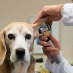
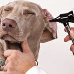
Your veterinarian will examine your dog’s ear canals and make sure there is nothing physically blocking the ear canal such as a tumor, polyp, or inflammation from chronic ear infections. However, as with congenital deafness, diagnosis of age-related hearing loss is difficult to do based on behavior. You may be able to get an idea if your dog may have hearing loss by snapping your fingers or clapping your hands behind your dog’s head. Your dog should startle or at least look in your direction, but they may also react if they sense the movement of air behind them, so it’s not a definitive test. The only way to definitively diagnose age-related hearing loss is with a BAER test, but with the limited number of testing centers available in the United States, the test is not considered to be practical for diagnosing age-related hearing loss. Instead, the presumptive diagnosis is based on the age of the dog, symptoms, and examination of the ears.
University of Cincinnati’s Facility for Education and Testing of Canine Hearing and Laboratory for Animal Bioacoustics, commonly referred to as FETCHLAB, has been working to develop hearing aids for dogs. As of 2019, nine dogs had been fitted for hearing aids, with varied success. The most common reasons for failure were the dogs not tolerating the hearing aids and inconsistent training in using the hearing aids once the dog was sent home. If you’re interested in trying to have your dog fitted for hearing aids due to age-related hearing loss, it may be beneficial to reach out to the FETCHLAB.
Once you determine that your dog has age-related hearing loss, if he’s not already trained to respond to hand signals, get started with training immediately while he still has some hearing. Starting to pair verbal commands with hand signals will make it easier for your dog to continue to follow commands using only the hand signals once his hearing is completely gone. Also start determining another way to get your dog’s attention when he’s not looking at you, such as stomping on the floor, using a flashlight, or releasing a scent that is appealing to him (such as opening a container of his favorite smelly treats). It will take some time to retrain your dog and retrain yourself, but being able to effectively communicate with your deaf dog will be worth the time and effort.
Ototoxicity
There are a number of drugs that have been identified as having the potential to cause damage to the inner ear. The damage can affect the cochlea (organ of hearing) or the vestibular system or both. It is difficult to determine the frequency of hearing loss as a consequence of using these drugs since dogs are able to compensate so readily to the loss of hearing and the differences in behavior are generally subtle. What little we do know is based on laboratory animal studies and human clinical studies. Of all the drugs with potential ototoxicity, the most commonly used in veterinary medicine are gentamicin, cisplatin, and furosemide.
Gentamicin is a broad-spectrum antibiotic commonly used in medications used to treat ear and eye infections. Hearing loss affects the higher frequencies first, as the hair cells responsible for high frequency hearing begin to die off. With long-term usage or high doses of gentamicin, the hair cells that are triggered by middle and low frequencies die off, the blood supply to the cochlea degenerates, and the auditory nerve is damaged and the hearing loss becomes complete. The hearing loss is irreversible, but recent studies in humans and laboratory animals indicate that the use of antioxidants help protect against hearing loss. However, studies of which antioxidants work best and at which dose in dogs have not been completed.
Cisplatin is a chemotherapy drug used primarily with sarcomas and carcinomas, such as osteosarcoma (bone cancer), transitional cell carcinoma (bladder cancer), and thryoid adenocarcinoma. Similarly to gentamicin, hearing loss affects the higher frequencies first before affecting the lower ranges, and the hearing loss is permanent. The severity of the deafness is directly related to the cumulative dose. In humans, up to 100% of patients have some degree of hearing loss with lengthy treatment protocols. Also like gentamicin, antioxidants seem to help prevent hearing loss, but there is some concern that antioxidants may also reduce the efficacy of cisplatin against cancer cells, so the use of antioxidants along with cisplatin is controversial.
Furosemide is a diuretic commonly used to treat edema (excess fluid collection in tissues) and hypertension (high blood pressure) in hospitalized patients. While the exact mechanism of toxicity is not fully understood, it’s suspected that the changes in electrolyte levels incurred by furosemide cause poor electrical conduction of the nerves involved in hearing. The hearing loss impacts the middle frequencies first, but the hearing loss is reversible once treatment with furosemide is stopped.
All of these drugs are important in veterinary medicine and the risk of hearing loss should not prevent their use, but steps can be taken to minimize the risk, such as not using the drug long-term or at high doses, or administering antioxidants along with the drug. Be sure to discuss the risks versus the benefits of these therapies with your veterinarian.
Noise-induced hearing loss
Just like in humans, dogs can have hearing loss caused by exposure to loud noises. Examples of dogs that might be affected include military working dogs exposed to combat situations, hunting dogs, and dogs housed in kennels with high noise levels. The middle ear has a protective mechanism in the form of muscles that contract the middle ear to reduce the amount of sound reaching the inner ear. However, those muscles can’t react quickly enough for sudden sounds such as gunfire, nor can they provide full protection against very loud sounds. Noise-related hearing loss is usually accompanied by age-related hearing loss, and there is usually a slow development of the hearing loss that goes unnoticed by owners until the hearing loss becomes significant enough that the dog is no longer able to fully compensate. The hearing loss can be temporary if the exposure is brief, but repeated or ongoing exposure to high levels of noise will result in permanent hearing loss.
There are a few companies that produce ear muffs for dogs, but unless they are exactly the right size and conformation for your dog’s head, they don’t provide the appropriate level of protection. The ear muffs are also quite bulky and may not be tolerated by your dog. For military working dogs, less bulky and more effective protective ear wear is being developed in conjunction with FETCHLAB, but the latest design is not available to the public at this point.
Otitis interna associated deafness in dogs
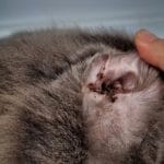
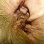
An infection of the inner ear is called otitis interna. Since the inner ear is deep within the skull and behind the eardrum, otitis interna is fairly rare, but is usually caused by chronic ear infections severe enough to damage the eardrum and permit bacteria to travel further down into the inner ear. Because mucus from the infection is present in the inner ear, the sound waves entering the ear are blocked, rendering the dog virtually deaf in that ear.
Signs of an inner ear infection include pawing or scratching at his ear, shaking his head, and possibly a reluctance to open his mouth and/or chew. Dogs with inner ear infections often have vestibular signs as well, since part of the system that helps a dog determine where their head and body is in space is located in the inner ear. Those signs can include a head tilt, leaning, falling, or rolling to the side, or walking in circles. Your dog may also be nauseated due to the dizziness he is feeling.
The facial nerve runs near the inner ear, so if it becomes damaged by the infection, you may see drooping of one side of the face, an inability to blink on the affected side, uneven pupil size, and/or elevation of the third eyelid. In particularly severe cases of inner ear infections that have crossed into the brain, a change in mentation (dull/depressed), neck pain, knuckling, difficulty walking, or seizure activity may be noted.
Definitive diagnosis of otitis interna requires the use of imaging techniques such as CT or MRI; both of which require general anesthesia in order to keep your dog perfectly still for the 20-45 minutes it takes to perform the imaging. If your dog has vestibular or neurologic signs, an MRI of the head is the most useful imaging technique. With an MRI, it can be determined if the infection has moved into the brain and to what extent. If the infection has indeed moved into the brain, a spinal tap is usually recommended and is performed immediately after the MRI while your pet is still under general anesthesia. The spinal tap can help confirm the presence of meningitis (inflammation of the lining of the brain and spinal cord due to infection). While advanced imaging is ideal, especially in cases where the dog has neurologic signs, a presumptive diagnosis of otitis interna is still possible with an examination of the ear, visualization of the eardrum, and the presence of mucus in or near the middle ear, if the dog permits it. Regardless of the method of diagnosis, an attempt will be made to collect the mucus present in the ear canal. The material will then be sent out to the laboratory for a culture and sensitivity test – this means the laboratory will attempt to grow any bacteria present in the sample and then test that bacteria against various antibiotics in order to determine the best antibiotic to use. This test takes 4-5 days to complete.
An inner ear infection is painful, so treatment will generally include oral pain medications such as an NSAID (non-steroidal antiinflammatory drug) and then an appropriate oral antibiotic. Your veterinarian may start your dog on a broad-spectrum antibiotic and then switch him to a different antibiotic if the culture and sensitivity test shows that a different antibiotic will be more effective. Antibiotic treatment should continue for at least three months for otitis interna cases, since antibiotics do not easily penetrate to the inner ear. If your dog has severe neurologic or vestibular symptoms, hospitalization for 24-48 hours may be recommended until the worst of the symptoms are alleviated; intravenous drugs and fluids can be provided to help keep him hydrated until he feels well enough to eat and drink on his own. Some neurologic signs such as a head tilt may never completely go away, but your dog’s ability to walk without falling over or leaning to the side should disappear fairly quickly once treatment is started, and his hearing should also return as the infection clears, assuming the treatment is started relatively soon after the symptoms start.
Final thoughts about deafness in dogs
The most common reasons for deafness in dogs is age-related hearing loss and congenital deafness. If vestibular or neurologic signs are ever noted in conjunction with hearing loss, it’s very important to get a diagnosis and start treatment promptly for an inner ear infection in order to prevent the infection from spreading to the brain, where it is much more difficult to treat. Most hearing loss in dogs is unfortunately irreversible, but most dogs are able to easily adapt to their hearing loss and make use of their other senses to compensate.