IN THIS POST
When a complete urinalysis is performed, one of the potential findings is crystals in your dog’s urine, or crystalluria. Most of the time, crystalluria is a normal finding in a dog; only rarely does it indicate something more serious. In fact, treatment of crystalluria is usually not necessary if no other unusual signs or symptoms are observed. Here we explore the underlying causes of crystals in dog urine along with the different types, and review the treatment methods (if needed) that are typically recommended.
How crystals in dog urine, or crystalluria is diagnosed:
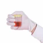
Your veterinarian will collect a urine sample from your dog. The reason for urine collection will determine the method of urine collection. If your veterinarian is simply screening the urine as part of an annual health check, then a free-catch method — catching the urine in a container as your dog voids their bladder — is sufficient. If your veterinarian is concerned about a urinary tract infection, then a procedure called cystocentesis is usually performed. Cystocentesis uses a long thin needle and syringe to collect urine directly from the bladder – this ensures the urine sample is not contaminated with any bacteria that might be present where the urine exits the dog, and therefore any bacteria found in the sample is the cause of the urinary tract infection.
Some veterinarians have the equipment to analyze the sample in house while others will need to send the sample out to the lab. In either case, the urine sample is spun down in a centrifuge. Any cells or crystals in the urine will be spun down to the bottom of the test tube while the liquid part of the urine stays on top. The material at the bottom of the test tube is collected, stained, and placed on a microscope slide. The sample is then examined under a microscope for the presence of bacteria, red or white blood cells, or crystals.
What causes crystalluria?
Crystals in dog urine usually form from minerals that are normally present in your dog’s urine. The presence of the crystals is determined by factors such as pH, temperature, and urine concentration (also known as urine specific gravity). Some crystals form in acidic urine (pH < 7.0) while others form in alkaline urine (pH > 7.0). Crystals also tend to form in urine that is more concentrated (higher specific gravity). Crystals can also form after the urine sample has been collected – the longer the urine sits before it is analyzed, the more crystals will form. Refrigeration of the urine sample will also cause crystals to form due to the decreased temperature causing the minerals to precipitate out of solution. Collection of the sample shortly after eating a meal may also cause more crystals to be present than usual.
However, some types of crystals will only form in response to specific medical conditions that may or may not be serious, and this is one of the reasons it is important to know the type of crystal that is present in your dog’s urine.
Types of crystals in dog urine:
Struvite (magnesium ammonium phosphate or triple phosphate)
Struvite crystals in dog urine may be a normal finding, and indeed, they are the most common type of crystal found in dog urine. Magnesium ammonium phosphate (struvite) normally remains dissolved in a dog’s urine as long as the pH of the urine is slightly acidic and the urine is not too concentrated. Therefore some dogs who normally have neutral or slightly alkaline urine, or are slightly dehydrated, may have struvite crystals; it’s simply not clinically significant for those dogs.
However, struvite crystals can also indicate the presence of struvite bladder stones, which is a complication of some urinary tract infections. Some bacteria that infect the bladder produce an enzyme called urease, which breaks down the urea that is normally present in a dog’s urine and produces ammonia. That ammonia then causes the urine to lose its acidity, allowing the formation of struvite crystals. Those struvite crystals can then collect around any cells or debris in the bladder and start growing into stones by collecting more and more struvite crystals. Struvite bladder stones are detected by either a radiograph or ultrasound. If stones (uroliths) are present in the bladder, treatment will be recommended; exactly which treatment will depend on the size of the stones and the risk of urinary tract obstruction.
If the stones are relatively small and there is little concern that they will cause a urinary tract obstruction, feeding a specialized diet to dissolve the stones may be recommended. These prescription diets will dissolve stones that are already present in the bladder and prevent new ones from forming. Dissolution of the stones can take anywhere from two weeks to three months. As the stones dissolve, they will release bacteria that have been trapped in the layers of the stone, potentially causing another infection, so your veterinarian will likely prescribe antibiotics during the dissolution process. Your dog’s progress will be monitored every four to six weeks with repeat radiographs and urinalyses.
Other possible non-surgical treatments for very small stones include a technique called urohydropropulsion. While your dog is under heavy sedation or general anesthesia, a special urinary catheter is passed into the bladder and the stones are flushed out. Stones could also possibly be removed using a cystoscope, which is an instrument with a camera and light source at the end; the cystoscope is passed into the bladder similar to a catheter. Tiny forceps are passed through the cystoscope, and using the camera for visualization, the veterinarian can grab the stones and remove them. Some specialty veterinary clinics may also offer ultrasonic dissolution, which is using high frequency ultrasound waves to break up the stones which can then be flushed out of the bladder.
For larger, multiple stones that are unlikely to respond to the dissolution diet or are likely to cause an obstruction, surgery to remove the stones will be recommended. While surgery is not to be taken lightly, it has the benefit of resolving the situation quickly, and once your dog has recovered from the procedure, they will no longer be in discomfort from the stones.
Dogs that do develop struvite bladder stones will likely need to be on a specialized diet for the rest of their lives. The recommended diet will be lower in phosphorus, magnesium, and protein, and also promote a more acidic urine pH. Since urinary tract infections (UTIs) are a major risk factor in developing these stones, dogs with signs of a UTI such as straining to urinate, frequent urination, blood in the urine, or urinating in unusual places should be checked out by a veterinarian as soon as possible.
Calcium Oxalate
The exact cause of calcium oxalate crystals in dog urine is complex and not well understood. However, like struvite crystals, calcium oxalate crystals may also be normal in some dogs, and tend to form in acidic and/or concentrated urine. Breeds prone to calcium oxalate crystals include miniature schnauzers, Yorkshire terriers, lhasa apsos, and miniature poodles.
Calcium oxalate crystals also tend to form with prolonged storage of the urine and refrigeration, so it’s important to consider under what conditions the urine sample was stored before it was analyzed. Diet may also be a consideration; diets that cause urine acidity (pH < 6.5) tend to predispose dogs to develop calcium oxalate crystals. The overuse of some antibiotics may also be a factor.
The presence of calcium oxalate crystals may also indicate the presence of bladder stones. Bladder stones are diagnosed by imaging the bladder with either radiography or ultrasound. Options for treatment are similar to struvite crystals, with the exception of using diet to dissolve the stones. Unfortunately, dissolution diets do not work with calcium oxalate stones. However, non-surgical options such as urohydropropulsion, cystoscopy, and ultrasonic dissolution may be available, and surgery to remove the stones is always an option. Once the stones have been removed, a change in diet can be made to help prevent recurrence, and increasing your dog’s water consumption (thereby diluting the urine) is another recommended strategy to prevent recurrence. Sadly, despite dietary changes and careful vigilance, calcium oxalate bladder stones are likely to recur.
A specific type of calcium oxalate crystal is found in the urine of dogs who ingested antifreeze, a toxin that causes difficulty walking, stupor, increased thirst and urination, and sudden and severe kidney damage. Therefore, urinalysis can be helpful in diagnosing suspected cases of antifreeze toxicity. Antifreeze ingestion is a medical emergency; the sooner treatment is started, the better the chance of survival. Treatment consists of intravenous fluid therapy and using drugs to prevent further absorption of the antifreeze. Once the kidneys start showing signs of damage at about 36 hours after ingestion, the prognosis becomes guarded to poor.
Ammonium urate (Ammonium biurate)
Ammonium urate crystals are often observed in the urine of dalmatians due to a genetic defect in a protein found in the kidneys. English bulldogs and Black Russian terriers are two other breeds in which the genetic defect has been identified. Otherwise, when these crystals are found in the urine of a dog of a different breed, it is a good indication of liver disease, particularly a condition called portosystemic shunt. Breeds at risk of a congenital (present from birth) portosystemic shunt include the Yorkshire terrier, schnauzer, Maltese, and pug.
The portal vein is a large vein that collects blood from the gastrointestinal tract, spleen, and pancreas and carries it to the liver. The liver would then remove toxins and bacteria from the blood before allowing it to enter circulation to the rest of the body (systemic circulation). A portosystemic shunt means that there is an abnormal connection between the portal vein and system circulation that bypasses (or shunts around) the liver altogether. When that happens, toxins enter systemic circulation and affect the brain and other tissues.
The most common clinical signs of a congenital portosystemic shunt include stunted growth, disorientation, staring into space, circling, head pressing, and seizures, so the condition is often diagnosed before the ammonium urate crystals are discovered. Sometimes the signs only occur after eating a high protein meal. Other dogs don’t show any signs until they are older and develop urinary problems such as kidney or bladder infections or ammonium urate stones.
Portosystemic shunts may also be acquired in older dogs as a result of liver disease. The signs are often related to the gastrointestinal tract, such as periodic episodes of bloody vomiting and diarrhea, increased thirst and urination, and fluid build-up in the abdomen. In these cases, the presence of ammonium urate crystals in the urine is helpful with diagnosing the condition.
Surgery to correct the congenital defect is usually recommended, particularly for dogs with neurologic signs. The surgery has a high success rate, with 95% of dogs returning to normal within 4-8 weeks after surgery. For dogs with minimal clinical signs or those with acquired shunts, medical management with diet is the usual treatment. Recommended diets include those with higher amounts of dairy-based or plant-based proteins in order to decrease the ammonium present in the bloodstream and urine. Canned food is used to increase water intake, which dilutes the urine, hopefully preventing crystal formation. Medical management in dogs with neurologic signs is not as successful, with over half of dogs being euthanized in less than a year due to progressive seizures and liver damage.
Cystine
Cystine crystals are present when an inherited kidney defect prevents some amino acids (protein building blocks) from being absorbed properly. This disorder affects many breeds of dogs, including dachshunds, English bulldogs, Scottish deerhounds, Newfoundlands, mastiffs, and miniature pinschers. It is suspected to be a sex-linked disorder, as almost all cases have been found in male dogs.
The presence of cystine crystals in the urine nearly always indicates the presence of cystine bladder stones. Treatment will depend on the clinical signs – in a dog that is having difficulty urinating or is obstructed (physically unable to urinate at all) – surgery to remove the stones from the bladder is usually recommended. Inability to urinate is a medical emergency – it is extremely uncomfortable and the risk of bladder rupture is high. If the dog doesn’t have any signs or symptoms, dietary management with canned food that promotes alkaline urine (higher pH) is recommended in order to prevent the formation or further growth of cystine stones.
Bilirubin
Bilirubin crystals are formed from the breakdown of red blood cells. Their presence in small numbers is normal in dogs, especially in highly concentrated urine samples from males. In the very rare times they are found in consistently high numbers in unconcentrated urine, they suggest a disease involving abnormal breakdown of red blood cells or liver disease. Your veterinarian will recommend further testing starting with a full blood work panel.
When is crystalluria a concern?
Crystalluria is often an incidental finding, and in many cases, no further investigation or treatment is needed; your veterinarian can advise you if this is the case with your dog. However, if your dog is showing signs of a urinary tract infection — having difficulty urinating, urinating frequently, urinating in unusual locations, or has blood in the urine — it’s important to have a veterinarian examine your dog and perform further diagnostic tests. The signs could be caused by bladder stones, and the earlier the stones are diagnosed, the more options may be available for treatment other than proceeding straight to surgical removal.
References
- bark & whiskers : Your Pet’s Biologically Imperfect Food Can Lead to Struvite Stones
- Pet Education: Bladder Stones (Urinary Calculi) in Dogs
- PetMD: Crystals in the Urine of Dogs
- The Whole Dog Journal: Canine Kidney Stone and Bladder Stone Prevention
- VCA Animal Hospitals: Struvite Bladder Stones in Dogs